ERCIM News 66July 2006Special theme: European Digital Library Contents This issue in pdf Subscription Archive: Next issue: October 2006 Next Special theme:
|
by Chris Kruszynski and Annette Kik
To conserve the Earth's coral reefs, biologists need to study them. In order to make better coral measurements possible, CWI and the Universiteit van Amsterdam developed sophisticated visualization methods to detect thickness, angles, lengths, spacing and branch ordering.
Coral reefs are important for the oceans' biodiversity and the growing nature-based tourism industry sector. They show a variety of forms depending on the environment, such as light, water flow and nutrients. Corals can, for instance, be ball-shaped or formed like a tree. The same species of coral can look completely different depending on environmental factors, which influence the growth pattern. To compare and classify specimens, it is important to make very precise measurements of coral thickness and branch distances. Biologists used to do this by hand, which takes a lot of time, and can cause unwanted errors. Due to the large number of branches, and complexity of corals, such manual measurements are generally only performed for a single metric - for example, branch length - or only for a small number of branches of a specimen.
To make these coral measurements easier, quicker, more accurate and robust, and more comprehensive, Krzysztof Kruszynski (CWI) and Jaap Kaandorp (Universiteit van Amsterdam) developed a method for the quantification of branching coral shape, for which coral specimens are scanned in a CT scanner. The scan data are filtered, segmented and transformed to a centerline skeleton. This method simplifies detection of features like branches, branching locations, and endpoints. The skeleton is then measured by the computer, and the results are subjected to statistical analysis; the measurements include thickness, angles, lengths, and spacing of branches.
However, several problems can occur. Noise comes from CT scanner ray scattering, decayed parts of the coral and creatures growing on it. Branches broken during transport are reattached with glue, influencing the measurements. It is difficult to fill holes - made by worms or human drilling - making it hard to distinguish the inside of the coral from the background. 'Skeleton loops' - due to low scanner resolution or branches growing back together - make branch ordering, and thus measurement, impossible. Noise filtering might affect the shape, but the noise itself influences the measurements, and must thus be reduced.
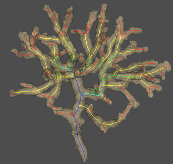 |
| Volume data from a CT scanner with a computed skeleton inside. Colours indicate branche numbers, counted from the outside. Picture CWI. |
Despite these problems, an interactive visualization system has been created and is being used. It is easier and quicker than older systems. The system has been created using existing open source software, such as the NLM Insight Segmentation and Registration Toolkit (ITK) for image processing, and the Visualization Toolkit (VTK) for visualization and interaction. New software was written for performing the actual measurements, using the VTK framework. The application area - coral biology - is novel for these techniques, some of which are quite advanced. For example, the Curvature Flow filter from ITK is an advanced image filtering technique which reduces the amount of noise with minimal impact on the shape of the coral.
Some of the tasks in the new system are performed manually. Segmentation - dividing the 3D image into objects of interest – is mostly automatic, but it produces hollow coral with holes in it. The inside is filled, and the holes are patched, but the results of this automatic process must be checked for correctness. The skeleton can be extracted without user intervention, but any loops in the skeleton must be disconnected by hand; there is no known technique to reliably determine where a part of the skeleton should be removed to disconnect the loop, or how much of the skeleton should actually be removed. The computer assists the user, and the number of loops is typically very small, making this an easy and quick task.
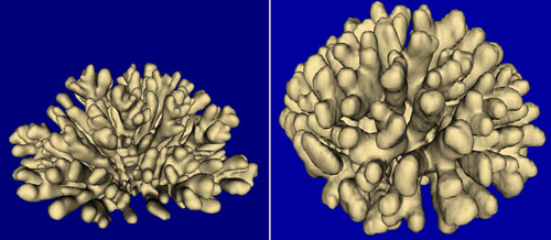 |
| Surface of a real, scanned coral and a simulated specimen. The right one is the real coral. Pictures CWI. |
In the future, the researchers want to create more advanced result visualizations; these are currently shown as a large collection of graphs and numbers. The accuracy of the computer measurements also still needs to be quantified. Clustering algorithms could detect similarities between species. Biologists can examine similarities and differences in shape, and study the correlation between shape and environment. This might help biologists with the important conservation challenges of coral reefs.
This work was partially carried out in the context of the Virtual Laboratory for e-Science project (http://www.vl-e.nl). This project is supported by a Bsik grant from the Dutch Ministry of Education, Culture and Science (OC&W) and is part of the ICT innovation program of the Ministry of Economic Affairs (EZ).
Links:
http://www.cwi.nl/ins3
http://www.science.uva.nl/research/scs/GF2004/
http://homepages.cwi.nl/~kruszyns/
Please contact:
Krzysztof Kruszynski, CWI
Tel: +31 20 592 4325
E-mail: K.J.Kruszynski![]() cwi.nl
cwi.nl