Extrinsic Registration of Heart Volume Images
by Mohamed El Ansari, Ilangko Balasingham, Tor A. Ramstad and Erik Fosse
Minimally invasive therapy has recently become one of the main areas of research in clinical medicine. The procedure is cost effective for both patients and hospitals. Advancements made in medical imaging have initiated the rapid development of minimally invasive surgery or key-hole surgery. The procedure is assisted by video-scopes. Its main drawback is manipulating the surgical fields through small incisions, which reduces direct vision and dexterity, and decreases the tactile feedback. It is hoped that enhanced vision through the addition of multimodal imageries will lead to a reduction in the difficulties faced by surgeons performing these procedures.
Minimally invasive heart surgery is a very useful technique for by-passing obstructed coronary arteries. The left internal mammary artery (LIMA) is a blood vessel located in the chest cavity, and in about 90 percent of all coronary bypass operations it constitutes the best available conduit for a surgical bypass to the major arteries of the heart. However, the soft tissue prevents the LIMA from being seen through the videoscope, and consequently it is difficult for the surgeon to localise it. By fusing vessel anatomy segmented from computed tomography (CT) angiography with the videoscopic images, this procedure can be carried out faster and more safely. The fusion can be done by means of registration, which consists of deriving a mapping transformation between the image space (CT) and the physical space, namely, the patient (see Figure 1).
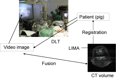 |
| The Direct Linear Transformation (DLT) is used to map physical space into video image space. By knowing the DLT and the registration between the CT image and physical space, we can fuse the CT and video images. |
Volume registration is a new area of research in medical processing and visulalisation. Registration of multimodal images makes it possible to combine different types of structural and functional information for diagnosis and surgical planning. Registration of images acquired with the same modality at different times or under different conditions allows the control of disease progression or regression. Registration of preoperative images with the physical space occupied by the patient during surgery is a fundamental step in image-guided heart surgery. It allows the fusion of the segmented LIMA from CT volume with endoscopic video images, thus enhancing the surgeon's field of view and the visualisation of the surgical anatomy.
Our approach is an extrinsic registration method, which is based on the implementation of spherical markers on the skin of the patient. A rigid-body transformation modelled as a rotation matrix and translation vector is considered. The registration approach is achieved in four steps:
- localisation of markers in CT images. A global thresholding method is applied to the CT images which produces binary images. The two morphological operations, erosion and dilatation, are applied to the binary images to remove small details and to fill whole appeared on the markers.
- localisation of markers in patient space. A stereoscopic method is used to compute the marker centroids in patient space
- correspondence between markers on image and physical spaces. The correspondence is achieved by using a heuristic measure of point pair affinity
- computation of the mapping transformation. The rotation matrix and the translation vector are computed by using a least squares method.
The method has been tested on synthesised data, and we hope to apply it to real data and exploit the results obtained to fuse the segmented LIMA from the CT volume into video images.
Mohammed El Ansari currently holds an ERCIM fellowship at NTNU.
Please contact:
Mohammed El Ansari, NTNU
E-mail: melansari@yahoo.com
|