
 This issue in
Analogic CNN Computing Fosters Detecting Stroke Signs
by Tamás Szabó and Péter Szolgay
Computationally intensive medical image processing applications typically require computational power usually not available with traditional computers. The CNN Applications Laboratory at the University of Veszprém, Hungary recently demonstrated analogic cellular neural network algorithms offering parallel cellular methods as well as wave methods for image enhancement and segmentation on computed tomography images.
In spite of the struggle against stroke, the frequency of cerebrovascular diseases which is ranked third in lethality worldwide has been increasing gradually all around the world. One of the most important reasons for the high lethality is the lack of efficient treatment within a critical period of three hours after the first observation of symptomes. As a result of fundamental and applied research we have developed a prototype of a cellular nonlinear network (CNN) based system with special analogic algorithms for two purposes. One is the enhancement of characteristics of diagnostic image features on computed tomography, CT images, and the other is the detection of stroke signs on CT images which can be misappreciated by a neuroradiologist. This system is integrated in a telemedical stroke expert network among hospitals and medical centers in Hungary.
A very high-speed computer architecture (four Tera equivalent floating point operation per second per square centimeter) with special algorithms reduces the computing time for a neuroradiologist to pre-process the images. A CNN may be embedded in a CT machine.
The Neurorad CNN System
CNN is a new computing paradigm of interdisciplinary interest, especially of two-dimensional biomedical imaging. The Neurorad CNN is a CNN-based system that is used to process CT scans. CNNs are parallel-computing, analog arrays, with very high computational speed. The cells are nonlinear dynamic processing elements. The connectivity among them is local in space. The program of the network is determined by the pattern of the weights of local interactions, the templates. The time-evolution of an analog transient represents the computation on a CNN. The CNN Universal Machine architecture invented in 1992, is a novel spatio-temporal array computer. The term analogic CNN computer is also used since analog spatio-temporal dynamics is combined with logic operations inbuilt in a stored programmable framework. Pixels of a CT image are mapped to the input of the cells. CT images have relatively low pixel and grayscale resolution. In the Neurorad CNN system several analogic algorithms are implemented.
Modification of the pixel-value distribution (the histogram) is done by differential equations, so the image contrast is enhanced while simultaneously reducing noise. This method can be improved for local contrast enhancement.
After separating the gray from the white matter, the average grayscale value of each hemispheres can be compared. Moreover, an active contour with initial position located on the central symmetry line of the CT image evolves to a final position, and its deformation is proportional to the quantified measurement of the hyperdense or hypodense areas of either of the two hemispheres.
The mathematical model we use for the segmentation of CT images is underlain by a nonlinear diffusion equation which is a partial differential equation, a PDE supplemented with reactive terms. Reactive terms are responsible for forcing the evolution-based process to stop at the edges that are unfortunately ill-defined in most of the CT images. This model - as being a more general model - displays the common qualitative characteristics of remarkable segmentation models.
A mask is constructed exactly by binary mathematical morphology for the segmented structures of interest. These structures are marked up afterwards in the original image. Later, erosion and dilation are used. The boundary of the mask is superimposed on the original image. Each result is verified by a medical expert. If no verification is gained, the process resumes to a new segmentation kernel.
Detecting Stroke Signs
One possible stroke sign, a hypodense region detected by a neuroradiologist and by the proposed system is shown in the figure.
|
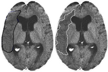
In the early hours, following stroke onset changes indicating the nature and extent of the stroke may be very subtle and difficult to see on a CT scan. To detect these early signs is important during future activities.
Please contact:
Tamás Szabó,
University of Veszprém, Hungary
Tel: +36 88 624799
E-mail: tszabo![]() almos.vein.hu
almos.vein.hu
Péter Szolgay, SZTAKI, Hungary
Tel: +36 1 2796128
E-mail: szolgay![]() sztaki.hu
sztaki.hu