Image Analysis by Scale-Space Operators
by Peter Johansen and Mads Nielsen
Over the last 5 years one of the key interests of the image analysis group at DIKU, Department of Computer Science, University of Copenhagen has been the analysis of images using scale-space operators. The aim has been to understand the multi-scale structure of images and thereby facilitate automatic or semi-automatic analysis of images. Tasks of interest are feature detection and segmentation and computation of stereo disparity and optic flow.At DIKU a group of approximately 5 researchers, lead by Prof. Peter Johansen is involved in this project. The primary collaborative partner is the 3D-Lab at the Medical Faculty, University of Copenhagen; where Mads Nielsen is coordinating the application of scale-space techniques to medical image analysis.
An image is digitally represented as intensity values on a pixel grid spaced at some resolution. By convolving this image with Gaussians of increasing width, a stack of images of gradually lower resolution than the original image is constructed. This is the scale-space. In this way image details are unconfounded from the pixel grid details, and the images at lower resolutions (higher scales) are differentiable making the toolbox of differential geometry available.
Convolving the image with Gaussians of increasing width causes the image to gradually simplify. For example, regions merge into large regions (splits, although rare, are also possible). In feature detection and image segmentation this simplification can be exploited by initially analysing at a resolution where the image is simple, and tracking the solution to a resolution where localisation is more precise and shapes more complex. This idea has been exploited in multi-scale image segmentation and in coarse-to-fine feature detection.
The local derivatives of the image at a given scale have been used for computation of the depth from a pair of stereo images, and also for computation of the local surface orientation and shape. This work is based upon analysis of how a deformation of the original image expresses itself in lower resolution images. This analysis also leads to a reformulation of earlier optic flow work in terms of scale-space.
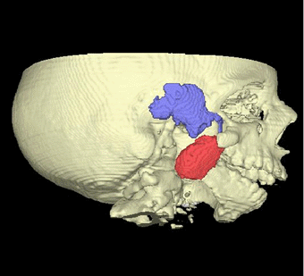
From a CT-scan the bone structure is easily identified from iso-intensity surfaces. Here also the muscular structure around the Jaw is identified using a multi-scale, watershed based, semi-automatic, 3D segmentation technique.The future direction of the project is to gain insight into the structural changes of images as scale varies and exploit this especially in the field of medical image analysis. This work is carried out in collaboration with 3D-Lab, Imaging Center Utrecht, Utrecht University Hospital, Royal Institute of Technology, Stockholm, and Department of Vision Sciences, University of Aston, Birmingham.
Please contact:
Peter Johansen - DIKU
Tel: +45 35 32 14 42
E-mail: peterjo@diku.dkMads Nielsen - 3D-Lab
Tel: +45 35 32 67 58
E-mail: malte@lab3d.odont.ku.dk