Mammogram and Echocardiogram Analysis by Using Cellular Neural Network Technology
by Akos Zarándy, Csaba Rekeczky, Márton Csapodi and Tamás Roska
Both mammogram and echocardiogram analysis require high performance image processing. In most cases an on-line analysis would demand computational power normally associated by super-computers, which is certainly not affordable in an ordinary clinical environment. Cellular Neural/ nonlinear Network (CNN) technology can answer to this challenge. CNN is a new, extremely high speed (Tera equivalent digital operations per second on a single chip), analog, programmable processor array. Due to its two-dimensional regular grid arrangement, the CNN can be efficiently implemented on silicon and it is well suited for image processing. There are already some working test chips which validate the paradigm. Encouraged by these results, in our mammogram and echocardiogram projects speed advantage of the CNN chips is being utilized. Our other objective is to design affordable hand-held devices to solve sophisticated on-line image analysis problems.
The work is being carried out at the Analogical and Neural Computing Systems Laboratory of SZTAKI, in cooperation with the National Institute of Oncology, Budapest and the State Hospital of Echocardiology, Balatonfüred.
Responsible for the projects at the medical institutions are Professor György Liszka and Ágota Petrányi (National Institute of Oncology) and Professor Ádám Tahy (State Hospital of Echocardiology).
Analogic Mammogram Diagnostic Workstation
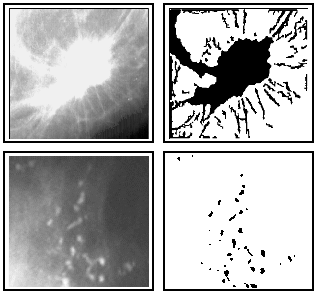
Diagnosing cancer tissues using X-ray mammograms is a time consuming task even for highly skilled radiologists because mammograms are low contrast, noisy images. In X-ray mammograms a breast cancer tumor is characterized by microcalcifications (small calcium carbonate particles), and tiny radial threads (spiculi) extending from the boundary of the tumor tissue. An Analogic Mammogram Diagnostic Workstation has been developed to help radiologists in their every day work. This workstation contains a Pentium PC for hosting all devices, an X-ray image scanner for digitizing mammograms, a CD writer providing for digital storage of acquired images and examination data, and a software package for image analysis and visualization, as well as for collecting the patient's personal and examination data. In the image processing part of the software package, two CNN analogic algorithms are employed (algorithms that combine analog and logic operations). The first algorithm finds and restores microcalcifications, while the second one finds the tiny spiculi around a given tumor kernel. All image processing algorithms are implemented via analogic CNN algorithms.
Figure 1: Upper image pair: an original mammogram image part and the detected spiculi around the tumor. Lower image pair: an original mammogram image part and the detected microcalcifications.At present, the CNN algorithms are executed by a digital emulator board which later will be replaced by analog VLSI CNN hardware when appropriate CNN chips become available. Analogic CNN technology will provide for a substantial improvement in the speed of computations. Our present system was introduced in clinical tests at the National Institute of Oncology, Budapest.
CNN Technology for Video-flow Processing of 2D Ultrasound Images
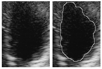
Feature extraction from echocardiographic images is of great importance for both quantitative and qualitative analysis of heart function. Such features include ventricular contours, myocardium thickness (the muscular substance of the heart), ventricular mass, etc. Since the major part of structural information is obtained from the location of the boundaries, most researchers carry out experiments using 2D ultrasound (B-mode) images and focus on contour estimation from a single frame or a sequence of frames. However, automatic processing of such images is very difficult due to poor quality (low signal-to-noise ratio) of echocardiographic images. Furthermore, a number of sophisticated methods implemented on conventional digital hardware platforms do not allow for an in vivo monitoring because of the prohibiting amount of computation required. The emerging new generation of CNN chips provides a true alternative in solving such challenging image processing problems. A CNN chip can be employed to process the flow of ultrasound images in real-time. We have synthesized a fuzzy-type analogic (analog and logic) CNN algorithm that is capable to detect the endocardial (inner) boundary of the left ventricle. In the preprocessing phase, adaptive image filtering and contrast enhancement are employed using constrained anisotropic diffusion. The task is to remove the so-called speckle noise due to coherent illumination and Rayleigh scattering caused by tissue microstructures. An iterative analogic algorithm is used that integrates region growing and object boundary detection to locate the central object (the left ventricle). The final result (the endocardial boundary) is extracted using morphological post processing. Figure 2 demonstrates the validity of the method superimposing the final output on the original image. The estimated time performance of the algorithm is around 100 microsec/line therefore the CNN chip can operate even at very high frame-rates (eg 64 frames/sec). Initial experiments indicate that ultrasonic imaging is well suited for the CNN computing paradigm. The CNN chip is capable of filtering and detecting, furthermore compressing and decompressing images without compromising the real-time nature of in vivo monitoring.
Figure 2: An example for image segmentation by using an analogic CNN algorithm: endocardial boundary detection in ultrasonic imaging. The original image of the left ventricle can be seen on the left, the final output (detected boundary) superimposed onto the original is on the right.
Please contact:
Akos Zarándy - SZTAKI
Tel: +361 269 8263
E-mail: zarandy@lutra.sztaki.huCsaba Rekeczky - SZTAKI
Tel: +361 269 8263
E-mail: rekeczky@lutra.sztaki.hu