
 ^
^
by Juhani Heinila
The work of medical doctors in radiology departments is changing rapidly because of the new digital imaging technology introduced into the hospitals. Although the diagnostic images on films still have many advantages, digital images displayed on the monitor and stored on digital archives (instead of huge hospital film archives) seem to be a reasonable and efficient solution in many cases. The change is twofold, because in addition to the new imaging technology, the way of making the diagnosis on the basis of digital images is different compared to the diagnosis based on films, and the image transfer network connecting specialists inside and between the hospitals causes changes on the level of human communication and team work between the medical doctors.
A teleradiology development project carried out at VTT produced a means for radiological image transfer. The results of the project were installed for clinical use in the Turku University Central Hospital (TUCH) in order to transfer X-ray images between two hospitals (30 kilometers apart).
Methods and design aspects
Unix operating system, the X Window System (X) and OSF-Motif were chosen for the software platform on the basis of the good networking properties of X (client-server model), which are essential in teleradiology. The images are transferred between workstations by FTP (File Transfer Protocol). The connection is based on an isolated 64 kbit/s point to point link. The maximum resolution of the digitizer is 3072*3072 pixels with 12 bit dynamics.
Two software packages were developed:
The graphical user interfaces (GUI) of the both workstations were designed to consist of three basic areas:
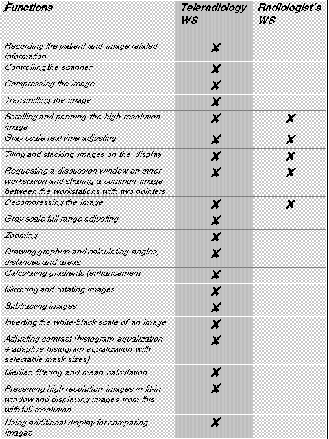
Features of the teleradiology system
In addition to the image presentation, there are many image related operations which should be available for diagnosing on radiologist's workstation. The teleradiology workstation could work with fewer image processing functions in our system. The most important functions of both workstations are summarized in the table.
The X client server network protocol allows a relatively easy way to produce functions characteristic for teleradiology, e.g., to opening windows on other workstations in the network, which could be used efficiently in opening a discussion window between the two workstations for writing text and sharing a common image with two separate pointers (one for each user).
Some of the filtering algorithms in radiologist's workstation have proved to be very useful in practice. One of the most fascinating is the adaptive histogram equalization (AHE). The power of AHE is the contrast enhancement of an image with minimum amount of noise enhancement.
Evaluation
The preliminary clinical evaluation of the teleradiology system has been carried out lately at TUCH. The evaluation was concentrating on comparing the diagnoses based on analogue (film) and digital image presentation in pulmonary diseases by using ROC curve techniques (ROC, Receiver Operating Characteristic). The results of the evaluations showed that for an experienced radiologist the 1 k x 1 k matrix was adequate in the detection of pneumothoraceas and fibrosis. On average, in the detection of pneumothoraceas, the difference was statistically significant between 1 k x 1 k digital image compared to analog radiographs. By using the resolution of 2 k x 2 k no such differences were found. In the detection of fibrosis no significant differences were found between analog films and the digital images presented on the monitor. The evaluation was based on individual performances of the specialists, although in practice the system supports co-operative work with common images on distant workstations.