
 This issue in
Development of a Real-Time 2D and 3D Echocardiographic Diagnostic System
by Dániel Hillier, Zsolt Czeilinger, András Vobornik, Zsolt Szálka, Gergely Soós, László Kék, Viktor Binzberger, David Lopez Vilarino and Csaba Rekeczky
Within the frame of a multi-disciplinary research project (TeleSense), the Analogic and Neural Computing Laboratory of SZTAKI has developed a prototype echocardiographic diagnostic system with telepresence capabilities. The system assists clinical diagnosis through novel three-dimensional reconstruction, display and two-dimensional analysis functionalities. These features significantly improve both the planning process of cardiac surgery and the efficiency of daily cardiac diagnosis.
Our research objective in the field of echocardiography was to create a prototype system in a PC environment that can efficiently support the daily routine of cardiologists. In the case of two-dimensional (2D) echocardiography, the aim was to develop a system having the ability to estimate in real time the volume of heart chambers, as well as analysing and evaluating the wall motion of the human heart. This information can be a valuable support for cardiologists making several medical diagnoses per day. In the case of three-dimensional (3D) echocardiography, the primary aim was to model and reconstruct the structure of interatrial muscles. These can give anatomical information that is useful in the preparation of successful surgery; typically the implantation of an occluder.
To achieve this aim, an experimental system based on a cellular neural network (CNN) was designed and implemented for efficient 2D/3D echocardiography image analysis and reconstruction. The 3D view of the human heart is reconstructed from 2D projections taken at different angles by an electronically controlled transducer. The analysis of these 2D slices (filtering, segmentation, contour tracking and content/context-based recognition) is designed as an analogic CNN-UM algorithm (UM stands for Universal Machine); the 3D reconstruction from the reduced data set (contour sub-sampling-interpolation, 3D rotation-translation, and polygonal reconstruction) is designed as a DSP algorithm. The system is based on the ACE-BOX computational infrastructure hosting both types of the aforementioned microprocessors, which ideally support these computationally demanding experiments.
The main hardware-software components of the ACE-BOX-based echocardiography diagnostic system have already been developed, and the algorithm optimization is an on-going effort. These experiments incorporate hundreds of video-flows of different patients, which are stored in a common database. Using a specially designed software tool, the cardiology specialists in our team support the experiments by semi-automatically tracking the borders of different chambers on all 2D projections. This way a set of 'groundtruth' data is created, which validates the performance of the segmentation and tracking algorithms and is used as a reference data set for the 3D reconstruction experiments.
|
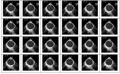
We have developed, implemented and tested three different versions of topographic cellular active contour techniques for heart chamber localization and tracking. These methods, called Constrained Wave Computing (CWC), Pixel Level Snakes (PLS) and Moving Patch Method (MPM), are solving the task of endocardial boundary tracking necessary for further processing. Figure 1 shows the output of the PLS algorithm in a single-chamber contour-tracking task that has been fully implemented on the ACE4k CNN-UM chip, meeting a video real-time performance with all system level overheads.
|
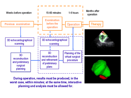
3D reconstruction examples are shown in Figure 2. In particular, two- and three-chamber analysis has been put in focus with the development of additional on-line tools that make it possible to provide efficient support to on-line surgical interventions. Figure 2 also summarizes the time criteria that must be met by engineering support in order to effectively help cardiac surgery. Screenshots illustrate the workflow when working with a 3D model of a reconstructed atrium.
One particularly valuable outcome of our work is a database that has been built with the help of the cooperating cardiologists and contains more than one thousand echocardiographic video-flows. More than half of the video-flows also contain reference contours that were traced manually by cardiologist experts. Our future work will include algorithm improvement and optimization based on this database and an experimental clinical introduction and testing of the entire system.
Our echocardiographic system also has native support for telepresence, that is, a technological environment that enables medical experts to see via wired or wireless network connection the results of a diagnosis undertaken at a remote site. At the same time the expert can consult the database and make use of the technological support developed for easy evaluation of clinical data
Diagnosis results can also be uploaded into the database. We have ensured that it is possible to reach the ACE-BOX analogical computing platform over TCP/IP; in other words, a cardiologist can access the hardware-software devices via a simple Internet connection, and use both our algorithms and the database.
The hospitals participating in the program (Saint Francis Hospital - SzFK, György Gottsegen National Institute for Cardiology - GGy-OKI) contributed by conducting specific medical experiments, continuously building the database, manually tracing reference contours and testing the user interfaces of developed algorithms. The design and integration efforts regarding the hardware architecture and the algorithmic framework were undertaken at SZTAKI. Péter Pázmány Catholic University (PPKE) and IT Consult-Pro Ltd. contributed by developing software tools, a simulation environment and core algorithms. A hardware-accelerating platform and connected software layers were implemented by AnaLogic Ltd GE Hungary Corp. provided hardware and software tools, support and user training.
We are confident that our system, capable of the above-mentioned functions, running with hardware acceleration and providing interactive support for 3D reconstruction and surgery planning, would be welcomed in medical institutions. In the current phase of the program, important developmental steps will be made towards a final market-ready product by conducting clinical introduction and testing.
Link:
http://lab.analogic.sztaki.hu/telesense
Please contact:
Csaba Rekeczky, SZTAKI, Hungary
Tel: +36 1 2796131
E-mail: rcsaba![]() sztaki.hu
sztaki.hu