
 This issue in
Virtual Tissue Matrix: A Pathologist Aid in Tissue Microarray Analysis
by Catherine M. Conway, Graham Dodrill, Darragh Lawler and Daniel G. O'Shea
Advancements in telepathology have resulted in the ability to review image of glass slides on-line. The development of Tissue Microarrays in the late 1990's, has significantly increased the throughput of biopsies analysed using immunohistochemistry, compared to more traditional methods. The Virtual Tissue Matrix was created to combine these two technologies, to allow virtual review of Tissue Microarray slides, and the recording of review data generated in a relational database.
Tissue Microarray's (TMA's) are glass slides, which permit simultaneous large-scale analysis of small tissue samples at DNA, RNA and protein expression. TMA's are manufactured by extracting cores, from donor tissue blocks. These cores are transferred into a TMA recipient block, which can house hundreds of cores from multiple donors. The TMA recipient blocks are then sectioned, and are transferred onto glass slides. One recipient block can create multiple glass slides, all of which have tissue spots arranged on the surface of the glass. The throughput of biopsy tissue reviewed, is greatly increased using TMA's, compared to more traditional methods.
TMA review involves manual examination of hundreds of tissue spots per slide, ranging in diameter from 0.6mm to 2mm per spot, using a conventional microscope. This process is labour intensive, and requires large amounts of time and concentration. There are a number of problems associated with glass slide reviews. Under a microscope, it is difficult to track which spot is under examination, at a given time. Pathologists can sometimes confuse their position on the slide, and record results associated with an incorrect spot. It is common practice for results to be handwritten at the time of the review, and subsequently entered into an Excel spreadsheet. Retrieval of results from these flat file spreadsheets can be difficult, due to the object oriented nature of the data. Technological developments in the area of telepathology have resulted in, reviews of histopathology slides being performed on line. This liberates the pathologist from the microscope, and also reduces the degree of human error associated with the scoring of Tissue MicroArrays using conventional microscopy.
|
Virtual Tissue Matrix (VTM) is a web application that allows, the review of TMA slides at remote locations. It consists of a user interface that provides images for review, and a relational database to store the generated results. The Medical Informatics Group, Dublin City University, first created the VTM in 2003.
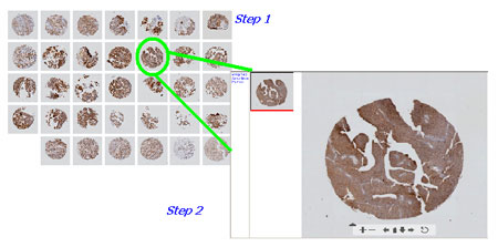
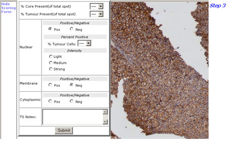
Once a user has successfully logged into the site, they are presented with a list of TMA recipient blocks available for review. On selecting one, a list of the associated TMA slides are presented. Once a TMA slide has been selected, an overview image of the slide appears. A larger image of each spot can be obtained by clicking on the image. The user can then scroll around the image, and change magnification as needed. Once a user is ready to record results for a spot, they click on an option button, which displays a pre-defined scoring form. The scoring page is a drop down form, which hovers over the image. It allows the user to record their results and submit the data to a relational database. The interface is designed to minimise data entry. The coordinating pathologist completes a form at the start of a study, which details the information they want to record. This information creates the pre-defined scoring form for each study.
The VTM was created using PHP, JavaScript, and an Oracle database. In order to allow the user to change the image magnification, the VTM uses a tool called Zoomifyer EZ. This allows high-resolution images to be viewed on a web page.
Future activities of the VTM will include the incorporation of an analysis package, which will assist in a pathologist interpretation of recorded data. Our aim is to have a tool that can run concurrently with the reviewing process. That will interpret trends and patterns in the results, and will highlight these areas of interest to the pathologists.
In order to validate the VTM, extensive testing of the system needs to take place. This will be done in conjunction with Beaumont Hospital Pathology Department and the Conway Institute, UCD.
Link:
http://www.telepathology.dcu.ie/VTM
To log into the site use the username
"ERCIM", password "ERCIM"
Please contact:
Catherine Conway, Dublin City University, Ireland
Tel: +353 1 5005281
Tel: catherine.conway3![]() mail.dcu.ie
mail.dcu.ie