|
ERCIM News No.48, January 2002 [contents]
|
|
|
|
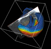
ICEMA: the Digital Heartby Frédérique Clément Making the best of the fantastic capabilities of the new cardiac imaging technologies, such as continuous 3D cardiac ultrasound, is the challenge taken up by INRIA through the ICEMA (Images of Cardiac Electro-Mechanical Activity) cooperative research initiative (ARC). ICEMA commenced in January 2000 and is developing into a skilled and multi-disciplinary initiative. Modelling experts, numerical analysts and image specialists are pooling their resources to design a fine numerical model of the activity of the heart, based on what is known of its functioning at the level of cardiac fibres. The originality of the researchers’ approach is to take into account both the electrical and the mechanical components of the organ activity. In effect, heart mechanical contractions are controlled by a conduction network issuing from the sinus node, the electrical depolarisation of which triggers muscular fibre contraction. Such an approach is not without difficulties, since it requires the coupling of two complex systems. It does, however, better conform to physiological models. Initially, project Sosso was able to describe the behaviour of the fibres that make up the myocardium by writing down a system of differential equations compatible with the molecular and cellular descriptions of contraction. Coupling this system to a model of the electrical activity of the heart – the control – makes it possible to locally describe the electromechanical activity. The global description takes into account geometrical aspects (the shape of the heart), dynamical aspects and the coupling with the other organs that take part in the system (blood volumes, vessels, etc.). The result is a system of partial differential equations developed by numerical analysts from projects Macs (INRIA Rocquencourt) and Sinus (INRIA Sophia Antipolis). These models are then tuned with the heart image reconstructions performed by the Epidaure research team at INRIA Sophia Antipolis, thereby obtaining a numerical description of the heart that is as realistic as possible. For their part, Epidaure researchers have reconstituted heart movements with the highest possible precision, based on 3D cardiac ultrasound image sequences supplied by Philips Medical Systems. For purposes of image segmentation, they have designed a volumetric model of the heart with an associated mechanical behaviour, which accounts for the heart’s very special torsional contraction. This model is in the process of being coupled with a reconstruction of the electrical field at the surface of the heart from electrocardiograms. The simulation of the mechanical behaviour of the heart thus obtained already provides satisfactory results. At the end of the initiative, one year from now, basic tools should be available that, once integrated into a platform, will allow clinicians to associate macroscopic perturbations visible on images or electrocardiograms with localised dysfunctions, either of muscular origin or related to the electrical control. The simulation aspect, which allows for prediction, could also be of interest in the study of surgical or pharmacological treatments. Link: Please contact: |